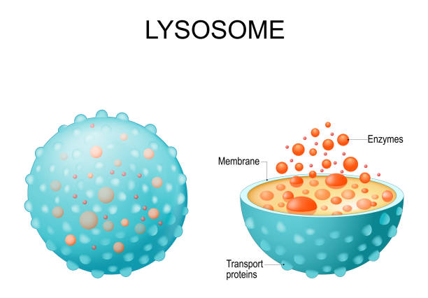
Lysosome. appearance, exterior and interior view. Cross section and Anatomy of the Lysosome: Hydrolytic enzymes, Membrane and transport proteins. Vector illustration
Browse 650+ lysosome stock photos and images available, or search for endoplasmic reticulum or golgi apparatus to find more great stock photos and pictures.
Lysosome. appearance, exterior and interior view. Cross section and Anatomy of the Lysosome: Hydrolytic enzymes, Membrane and transport proteins. Vector illustration
Anatomy of the Lysosome: Hydrolytic enzymes, Membrane and transport proteins. This organelle use the enzymes to break down and digest food particles, engulfed viruses or bacteria in the cell. Vector diagram for medical use

autophagy of mitochondria. Diagram of the process of autophagy from Forming a membrane and autophagosome to fuse phagosome and lysosome when contents of the vesicles are degraded and recycled. Autophagy defects linked to various diseases and cancer development. Vector poster
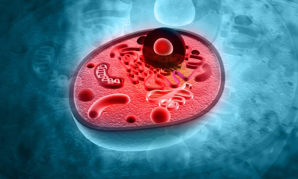
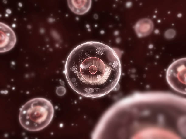
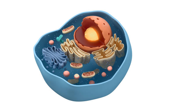
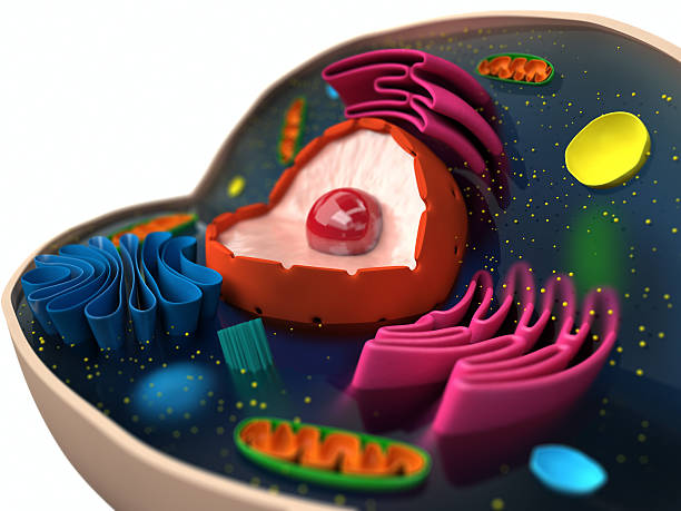
Cell Structure on blue background. 3d illustration

Endocytosis. The transport of macromolecules into a cell in a vesicle. Vector illustration design

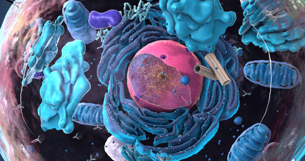
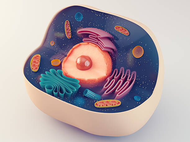
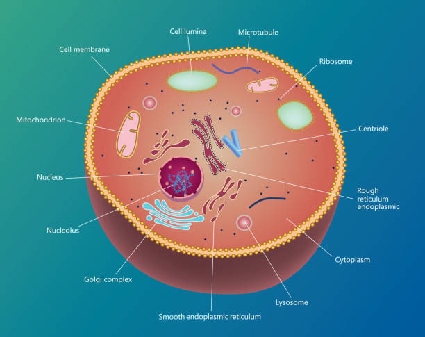
Structure of human cell, anatomy of cell, cellular environment, cellular concept with organelle: nucleus, membrane, mitochondria, Golgi apparatus 3d rendering
Anatomical structure of biological animal cell with organelles

Autophagy process. From forms a double membrane and autophagosome formation to Autophagosome fuses with a lysosome and Degradation of cellular component. Cell recycling. Cancer therapy and Immune regulation. Cell organelles. vector illustration isolated on white background.

3d render of eukaryotic cells
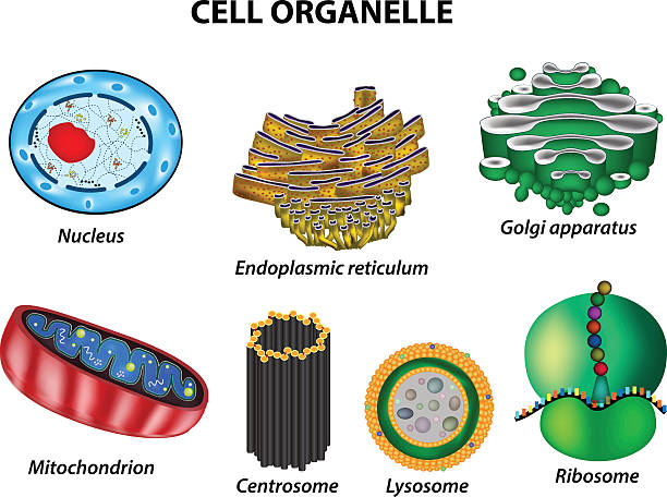
Set the cell organelles. Nucleus, endoplasmic reticulum, Golgi apparatus, mitochondria, centrosome, lysosome, the ribosome. Infographics Vector illustration on isolated background

Endosomes - 3d rendered image of Endosomes are membrane-bound organelles. They are parts of endocytic membrane transport pathway. Endomembrane system in cells. Bilayer Lipid Membrane. Abstract medical illustration. Microscopic view. Microbiologic research concept.
Gradient and transparent effect used.
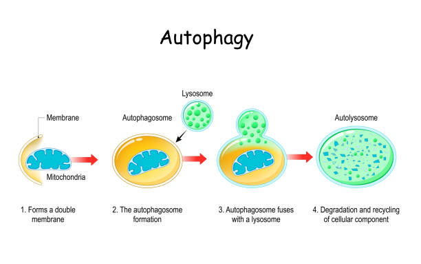
Autophagy steps. Schematic diagram. Natural mechanism in the cell that removes unnecessary components. Vector illustration

Autophagy. Computer illustration of a lysosome (orange) fusing with an autophagosome (large sphere). Autophagy (autophagocytosis) is the natural mechanism that destroys unnecessary or dysfunctional cellular components and recycles their materials. The target components are first isolated from the rest of the cell within the double-membraned autophagosome. This then fuses with a lysosome, the contents of which degrade the target components. The 2016 Nobel Prize in Physiology or Medicine was awarded to Japanese cell biologist Yoshinori Ohsumi for his discoveries of mechanisms for autophagy.

Endosomes - 3d rendered image of Endosomes are membrane-bound organelles. They are parts of endocytic membrane transport pathway. Endomembrane system in cells. Bilayer Lipid Membrane. Abstract medical illustration. Microscopic view. Microbiologic research concept.

Autophagy is a process where cells degrade and recycle their own components. Damaged or unnecessary cellular material is enclosed in a membrane, forming an autophagosome, which then fuses with a lysosome. The lysosome contains enzymes that break down the materials for recycling. It is a work created by CG, not created by AI. It was designed and created by myself.

lysosome anatomy. Hydrolytic enzymes, Membrane and transport proteins. This organelle use the enzymes to break-down virus particles or bacteria in phagocytosis of macrophages. Vector illustration

Structure of Tissue of Spleen Human, Liver Human and Kidney Human under the microscope in Lab.

Illustration of animal cell anatomy

Endosomes - 3d rendered image of Endosomes are membrane-bound organelles. They are parts of endocytic membrane transport pathway. Endomembrane system in cells. Bilayer Lipid Membrane. Abstract medical illustration. Microscopic view. Microbiologic research concept.

Human cells anatomy blue color. 3d illustration
Gradient and transparent effect used.
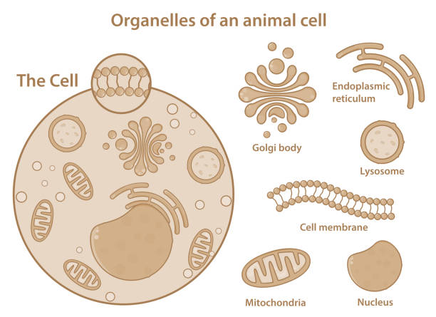
Organelles of an animal cell showing different components present in a eukaryotic cell. Simple and clear medical illustration. Major parts of a cell only. Aesthetic graphics.

Functions of lysosomes with anatomical explanation outline collection set. Educational labeled organelle with hydrolytic enzymes vector illustration. List with gene, plasma and cholesterol regulation.
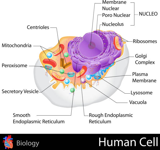
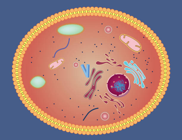
easy to edit vector illustration of human cell structure

Human cell structure: nucleus, cytoplasm, golgi, organelles mitochondria, ribosomes, lysosomes, cell membrane. Genetic DNA replication, RNA, protein synthesis, biology medical science. 3D cell anatomy scientific illustration

Numerous intracellular organelles are often covered by lipid bilayers. The vesicle is split in half and its internal structure is imaged. The bilayer is made up of small lipid monomers facing each other.
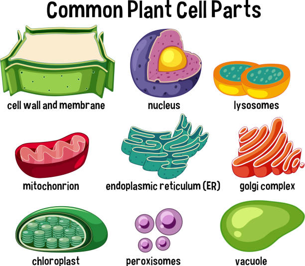
Common plant cell parts illustration

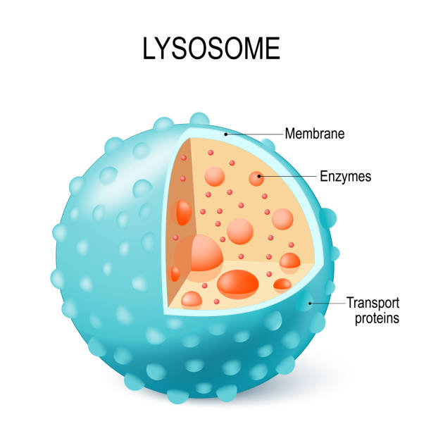
Anatomy of the Lysosome: Hydrolytic enzymes, Membrane and transport proteins vector illustration on white background
Internal structure of an animal cell, 3d rendering. Section view. Computer digital drawing.
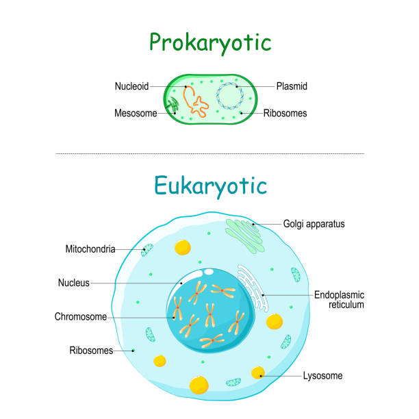
Prokaryote vs Eukaryote. illustration of eukaryotic and prokaryotic cell with text. Differences between Prokaryotic and Eukaryotic cells. vector diagram for education, medical, biological and science use

Lysosome. Anatomy of the Lysosome: Hydrolytic enzymes, Membrane and transport proteins. organelle use the enzymes to break down and digest food particles, engulfed viruses or bacteria in the cell. Vector illustration
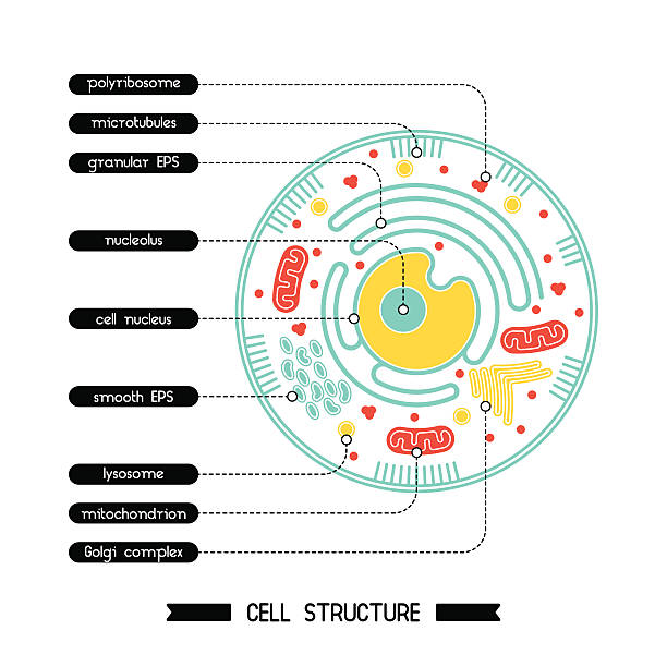
Isolated cell biology pictogram. Cell anatomy structure vector illustration. Cell structure detailed colorful anatomy with description.

human cell components of an eukaryotic cell, nucleus and organelles and plasma membrane Golgi, mitochondria, smooth ER, rough ER, lysosome, endosome, 3d rendering

Anatomy of the Lysosome: Hydrolytic enzymes, Membrane and transport proteins. This organelle use the enzymes to break down and digest food particles, engulfed viruses or bacteria in the cell. Vector diagram for medical use
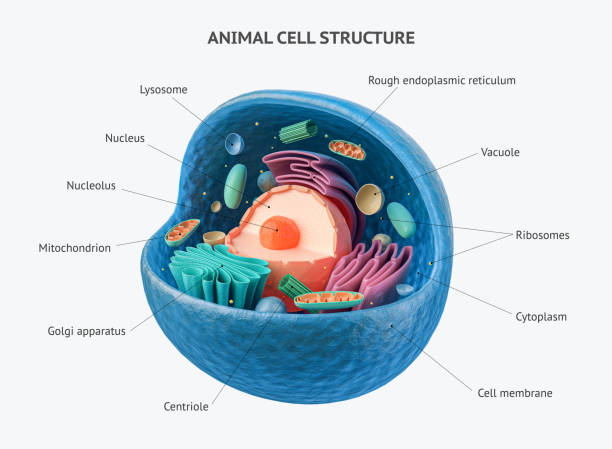
3d rendering of biological animal cell with organelles cross section isolated on white. Animal cell with placed text annotations to all organelles

Apoptosis (chromosome condensation, nuclear fragmentation), autophagy (autophagosome formation), necrosis (membrane rupture, organelles swelling)

Movement through the Plasma Membrane methods

Internal structure of an animal cell, 3d rendering. Section view. Computer digital drawing.

Illustration showing the internal structure of the human cell with the cytoplasmic membrane, nucleus and organelles isolated on white

Isolated multi lamellar vesicle or multilayered liposome in the black background 3d rendering

The different types of endocytosis: receptor-mediated endocytosis, pinocytosis (cell drinking) and phagocytosis (cell eating). vesicle, coated vesicle, and phagosome. vector illustration for medical, educational and science use

Bilayer Lipid Membrane - 3d rendered image of Cell Membrane Phospholipid Structure, Phospholipid bilayer of the cell membrane. Abstract medical illustration. Microscopic view. Microbiologic research concept.
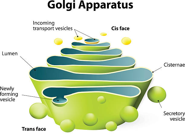
Golgi apparatus. Golgi Complex plays an important role in the modification and transport of proteins within the cell
The structure of a biological cell (macro)

Detailed process of phagocytosis in four stages: entrapment of food particle, formation of food vacuole within cell, fusion of vacuole and lysosomes, digestion of food particle.

Phagocytosis. macrophage absorption of bacteria. Stages of mechanism of the immune response from entrapment or endocytosis to phagosome formation, degradation and exocytosis. Vector illustration. Medical poster.

Endocytosis. Lysosome digesting food. Part of cell (plasma membrane, cytoplasm and lysosome), with food vacuole. Lysosome fusing with the food vacuole. Vector illustration

The macrophage is a large white blood cell that is an integral part of our immune system. Its job is to locate microscopic foreign bodies and 'eat' them. Macrophages use the process of phagocytosis to engulf particles and then digest them. Some macrophages roam the body and some stay in one particular area